The control centers for most nervous system functions are located in the brain. The cerebrum, cerebellum, diencephalon, and brain stem control specialized groups of functions. The brain modulates the spinal cord, which is primarily a reflex response center. Most simple reflexes result from neurotransmission through the reflex arc, a three-neuron chain composed of sensory, connecting and motor neurons. The cerebrum, which controls all advanced mental activities, is divided into two hemispheres; the corpus callosum, a connecting bridge of nerve fibers, joins the hemispheres. Large fissures divide the two hemispheres into the frontal, parietal, temporal, and occipital lobes. The cerebral cortex, which covers the surface of each hemisphere, is composed of gray matter containing the cell bodies of neurons and white matter containing their myelinated nerve fibers. Masses of gray matter called basal ganglia are found deep within each cerebral hemisphere; basal ganglia form a part of extrapyramidal system (2007). This controls the coordination of muscle groups that function together to perform voluntary motion. The internal capsule is white matter, consisting of bundles of nerve fibers, that passes through the basal ganglia, carrying sensory and motor impulses to and from the cerebral cortex. Parts of the cortex called functional areas are related to specialized functions; however, because these areas are extensively interconnected. The motor area controls voluntary motor activity. This area contains neurons that control specific body parts. Neurons in the upper part of the motor area control muscles in the lower part of the body. Neurons in the lower part of the area control muscles in the head, neck, and upper body. The number of neurons supplying a muscle depends on the type of movement it performs. Muscle capable of fine movements, such as finger muscles, have large areas of cortical representation. Muscles capable only of relatively gross movements, such as large limb and back muscles, are represented with smaller areas. The sensory area receives sensory sensory impulses. Each cerebral hemisphere receives sensory impulses from, or supplies motor impulses to the opposite side of the body because almost all of the fiber tracts - bundles of nerve fiber that carry impulses - cross to the opposite side of the brain, brain stem, or spinal cord as they ascend and descend the CNS (2007). Cortical areas associated with vision are located in the occipital lobe; those associated with hearing are located with hearing are located in the temporal lobes. Motor areas related to speech are located in the frontal lobes; cortical areas related to olfactory sensations are on the under surface of the temporal lobes. Other cortical areas surrounding the primary areas are called association areas although they're concerned with the same types of functions as the primary areas, they're more involved with interpretation, learning, and memory. The frontal areas primarily involve personality and judgement. "The cerebellum receives sensory impulses from muscles, joints, and tendons that convey a sense of position; it also receives impulses regulate muscle groups that coordinate position and balance. Bundles of fiber called the cerebellar peduncles connect the cerebellum to the brain stem" (Mareib, 2009). Cerebellar peduncles transmit nerve impulses to the spinal cord, medulla, and brain. The peduncles transmit impulses from the cerebellum to the thalamus for eventual transmission to the cortex. The diencephalon connects to the top of the brain stem and contains the third ventricle, the ventricle being a hollow part or cavity in an organ. The thalamus, which forms the lateral walls of the third ventricle, contains relay stations that receive sensory impulses and transmit them to the cortex. The hypothalamus contains neurons that control hormone output from the endocrine glands (2007). These neurons control basic body functions such as temperature regulation and food intake. The brains stem is divided into the midbrain, pons, and medulla. The midbrain contains cell bodies of cranial nerves and large nerve fiber bundles that convey impulses to and from cerebral hemispheres. The pons is a transverse bridge of fibers tracts extending to the cerebellum. Finally, the medulla is the lowest part of the brain stem; it forms the floor of the fourth ventricle, which is covered by the cerebellum.
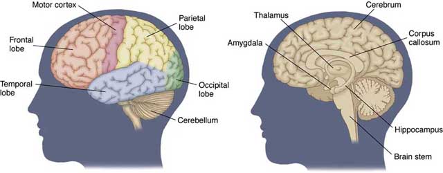
The anatomy of the human brain, exterior view showing the lobes (left) and the interior sections (right). (World of Anatomy and Physiology, 2010)
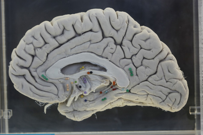
A picture depicting a real human brain. (Gale Science in Context, 2011)
